
This page showcases creative teaching and teaching excellence of our faculty members.
Digital denture set-up and iDDS: Very positive feedback from students
Drs Ting-Ling Chang, Takahiro Ogawa, Daniela Orellana, Denny Chao, Ben Wu
Digital denture set-up project implemented to Immediate Denture Course (RFE221) was evaluated by the course chairs from the students’ perspective. The result of students’ survey was extremely positive for this new implementation, faculty efforts, and usage of iDDS resources. The comments include:

a very cool experience

This was a very cool experience. I was able to complete the setup and achieve contact in less than two hours, which is a much more speedy process than doing this by hand. I wonder how accurate it is to transfer this digital setup to a physical one. We did not get to experience that transition. If that process works out seamlessly, then this digital alternative is a more than ideal replacement to creating a setup on a physical cast.
The immediate denture project gave me a clinical picture towards the whole project.
I thought overall the digital aspect was a really nice move towards modernizing the preclinical aspect and certain elements worked really well (like the digital MAP, scanning, and uploading our scans). However, I think there could be better improvement to the software (specifically the adjusting the contacts in the digital immediates tooth set up).
I think it's great that digital dentistry is being included into the lab sessions. The only improvement I can think of is allowing students to have more time to play around with the software.
Implementing digital technology for Maxillofacial Prosthetics fellows
Dr. Denny Chao
Since joining the Division of Advanced Prosthodontics at UCLA in 2019, Dr. Chao has implemented the use of various digital technology to teach and treat patients at the Maxillofacial Prosthetics clinic. Dr. Chao is present chairside and instructs Maxillofacial Prosthetics Fellows in the proper use of intraoral structured-light 3D scanners and facial scanners. He created step-by-step PowerPoint instructions in the virtual planning of dental and zygomatic implants in maxillectomy patients as well as in the digital design of guided zygomatic implant templates, titanium clasp assemblies, and facial prostheses. Additionally, he is in the process of converting the step-by-step instructions into video format. Dr. Chao is also versed in the computer-aided manufacturing process of digitally designed parts and educates the fellows on the operation and maintenance of 3D printers and milling machines. Dr. Chao disseminates knowledge regarding digital technology in dentistry with the goal of executing procedures faster, cheaper, and better.
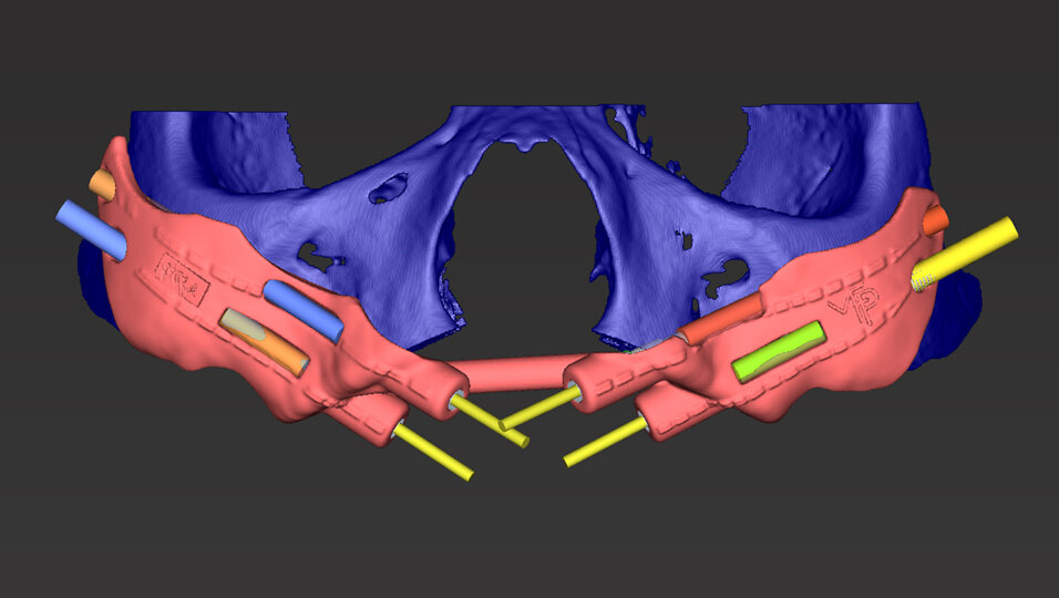
The digital design of a guided zygomatic implant template for the placement of 4 zygomatic implants in a patient undergoing bilateral maxillectomy.


The digital design of clasp assemblies for an immediate surgical obturator for a patient undergoing maxillectomy. The clasp assemblies are manufactured out of titanium with computer numerical control (CNC) machining.

A facial scan is obtained with a structured-light 3D scanner in preparation for the computer-aided design of a left auricular prosthesis.

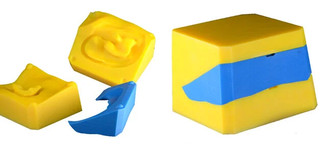

The auricular prosthesis is designed and digitally festooned (texturized). A 3-part processing mold is designed and 3D printed. Silicone will be utilized inside of this mold to make the actual auricular prosthesis. The finished left auricular prosthesis.
Digital design and mouth-prep evaluation implemented to RPD
Drs Daniela Orellana and Ting-Ling Chang

Traditionally, we have obtained diagnostic cast through conventional methods which includes alginate impression and stone cast. Recognizing the importance to adopt and teach digital innovations, we’ve enhanced our predoctoral curriculum to include projects which students become familiar with these cutting-edge technologies. Removable partial denture courses (RFE219 and 220).

We introduced intraoral scanners as an alternative to obtain a diagnostic cast. In addition, from this 3D moles, we have taught students how to manipulate to find an ideal path of insertion for a rigid removable partial denture prosthesis. This step has always been challenging for students to grasp, but now using digital technology that aids in the diagnosis and design, students can magnify the high-resolution scans and evaluate how minor movements affects the future path of insertion of the prosthesis.


We also created standardized teeth preparations and scanned them for students to be able to visualize ideal contours and reductions. The STL files of the ideal tooth preparation was uploaded in the COMPARE software. After each student did their laboratory project, they can scan their tooth preparation and this software allows to compare it to a known standardized preparation. Actual numerical values are generated by the software, making a more objective and reliable feedback for our students.
We did the same for other procedures such as rest preparations for the removable partial dentures. Standardized rest preparations were recorded using an intraoral scanner and imported in the software. Students can scan their own preparations and do a virtual comparison between their sample and the ideal contours.
Dental Material Course customized for residents
From clinical decision-making to research ideas
Dr. Alireza Moshaverinia
Dr. Moshaverinia is directing an Advanced Dental Material Course for Graduate Prosthodontics residents. This course is designed to provide the graduate student with fundamental and in-depth knowledge of the properties, selection criteria, and proper use of dental materials, beyond that possible in the predoctoral dental curriculum. The graduate student should also be able to discuss research publications on these materials in the dental literature, describe the general experimental procedures for determining the properties of these materials, and make informed decisions about materials selection.

OSCE developed and implemented during the pandemic
Drs Ting-Ling Chang, Daniela Orellana, and Takahiro Ogawa
Drs Chang, Orellana, and Ogawa developed an OSCE (Objective Structured Clinical Exams) in removable partial dentures (RPD) and immediate dentures (ID). This is a part of teaching modification and improvement during the COVID-19 pandemic and to ensure a valid, objective, and reliable assessment for clinical competencies of students. Drs Chang, Orellana, and Ogawa created a comprehensive online OSCE from the limited resources and time available during the pandemic to provide quality assessment of students’ skills and knowledge during the limited usage of clinic and patient care.

Implementing digital set-up
Drs Takahiro Ogawa and Denny Chao
Dr. Ogawa and Chao developed and implemented a digital denture teeth set-up project in the preclinical course of Immediate Denture (RFE306). This allows students to learn and experience digital tooth extraction, digital survey of the alveolar anatomy, digital set-up of denture teeth, digital analysis of occlusion using a virtual articulator, digital festooning, and overall work-flow of making an immediate denture using digital techniques. Dr Ogawa created a step-by-step video instruction by screen-capturing the digital software that entirely covers from the scanning, data transfer, software operation, necessary digital tools, and information and knowledge required to interpret the work. The project was conducted in the iDDS room and students completed the project by submitting screen capture images of the set-up and articulation.


